COLUMBIA | Zuckerman Institute

Polleux Lab


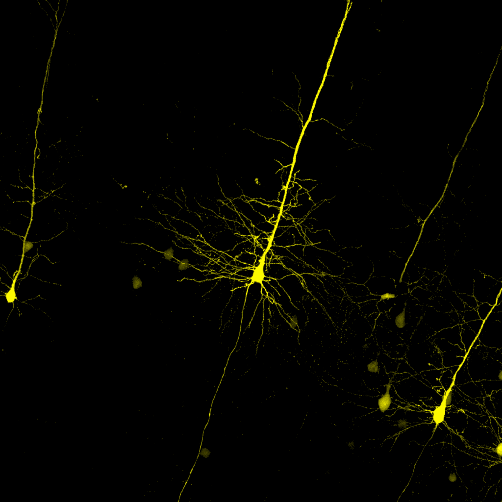
Circuit development, evolution and maintenance

Research

Research in the Polleux laboratory focuses on three important questions relevant to the brain

Cell biology of the neuron
What are the cellular and molecular mechanisms patterning the connectivity of cortical circuits during mammalian development?

Evolution
What are the genetic mechanisms that led to the evolution of human cortical circuits?

Aging
What are the signaling mechanisms underlying synaptic loss during early stages of Alzheimer’s Disease?

Our work provides new insights into the cellular and molecular mechanisms underlying the establishment and maintenance of brain connectivity and has significant implications for our understanding of the pathophysiological mechanisms underlying socially-devastating neurodevelopmental disorders and neurodegenerative diseases.

About the PI

Dr. Franck Polleux did his undergraduate and graduate studies at Université Claude Bernard in Lyon, (France) where he obtained his Ph.D. in Neuroscience in 1997. He then joined the laboratory of Dr. Anirvan Ghosh at Johns Hopkins University for his post-doctoral training.

In 2002, Dr Polleux was hired as an Assistant Professor in the Neuroscience Center and Department of Pharmacology at the University of North Carolina- Chapel Hill where he became an Associate Professor in 2008. In August 2010, he joined The Scripps Research Institute in La Jolla, California. In November 2013, he was recruited as a Professor in the Department of Neuroscience at Columbia University to join the new Mortimer B. Zuckerman Mind, Brain, Behavior Institute.

Throughout his career, Dr Polleux has focused on the identification of the molecular mechanisms underlying neuronal development in the mammalian brain. More recently, his lab started studying the genetic basis of human brain evolution as well as the signaling pathways underlying synaptic loss during early stages of Alzheimer’s Disease progression.

Watch Franck's talk on World Wide Neuro[CV]

The Team

Daniel Virga (he/him)
Graduate Student

Stevie Hamilton (she/her)
Graduate Student
Kevin Gonzalez
Graduate Student

Sergio Bernal-Garcia (he/him)
Graduate Student
Victoria L. Hewitt (she/her)
PostDoc

Justin O'Hare
PostDoc

Eugenie Peze-Heidsieck (she/her)
PostDoc

Martina Proietti Onori (she/her)
PostDoc
Carlos Diaz Salazar Albeda
PostDoc

Qiaolian Liu
Research Technician

Rhythm Sharma (she/her)
Lab Manager

A sneak peak at some of our Tools

In Utero Electroporation and Neuron Reconstruction

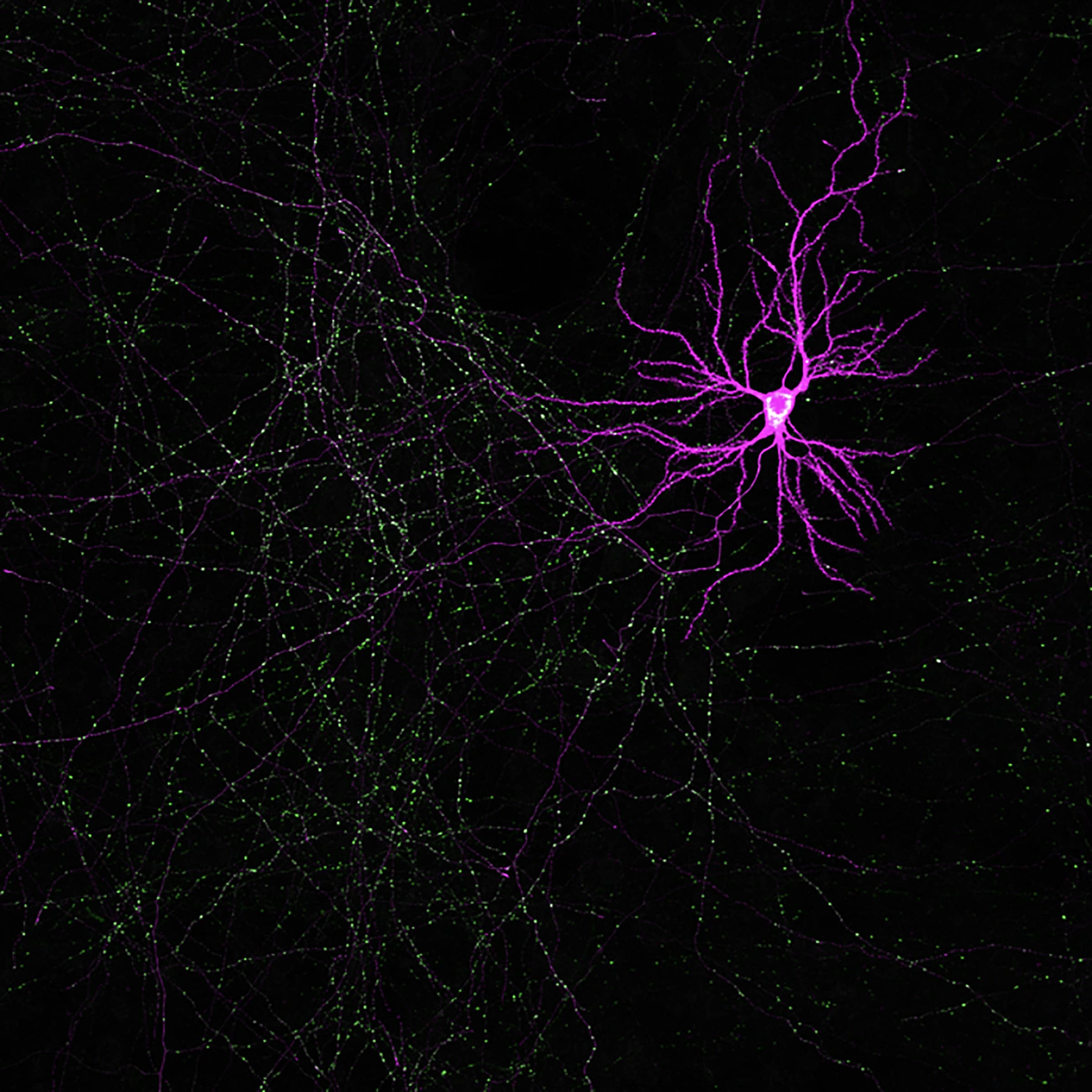
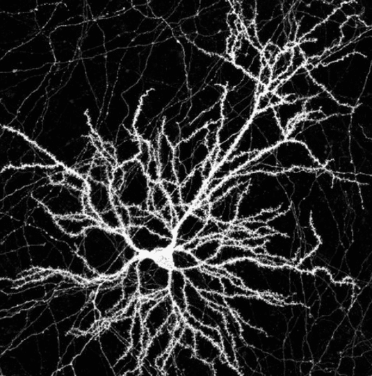
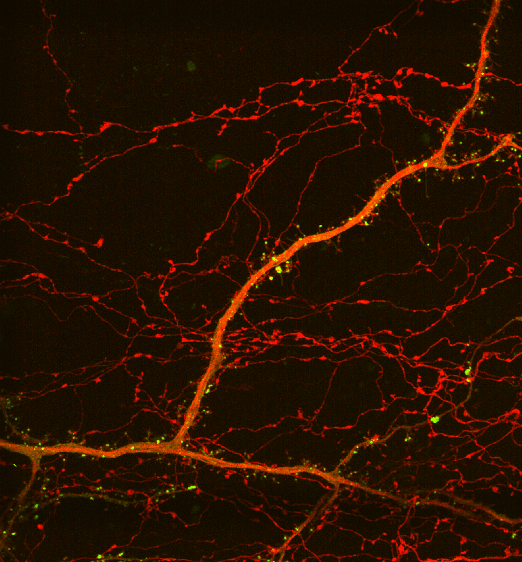
We developed a new, open-source, reconstruction platform for mapping the size and spatial distribution of E and I synapses received by individual, genetically-labeled, layer 2/3 cortical pyramidal neurons (PNs) in vivo. We mapped over 90,000 E and I synapses across twelve L2/3 PNs and uncovered structured organization of E and I synapses across dendritic domains as well as within individual dendritic segments in these cells.
The image describes the workflow for excitatory and inhibitory synaptic reconstruction across whole neurons with Vaa3D Synapse Detector program developed in collaboration with the Allen Institute for Brain Science.
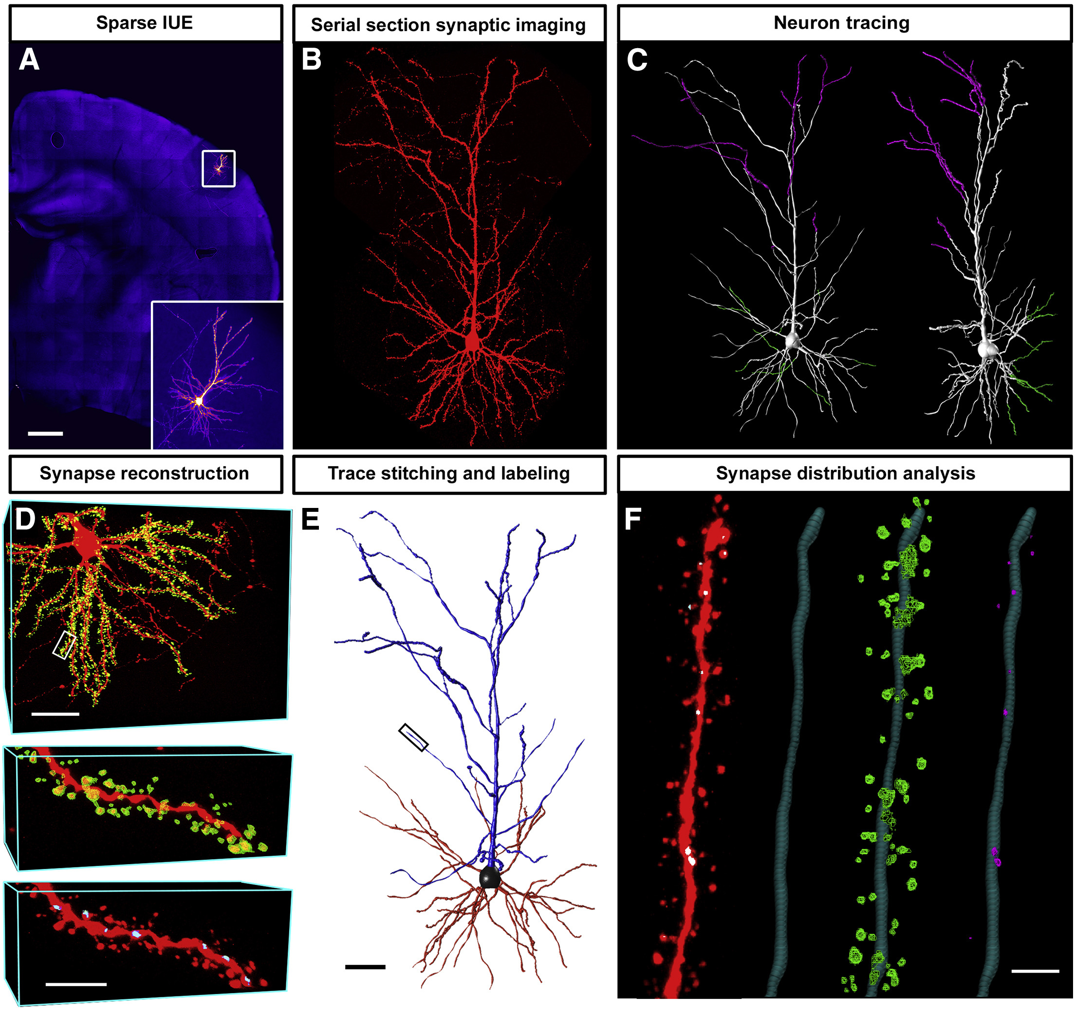
This work is featured in this paper by Daniel Iascone

Rabies Tracing and Neural Connectivity Reconstruction
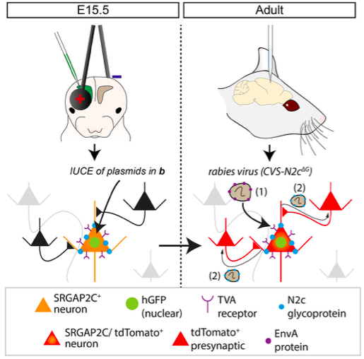
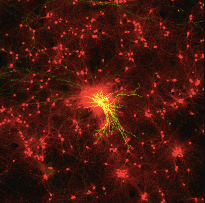
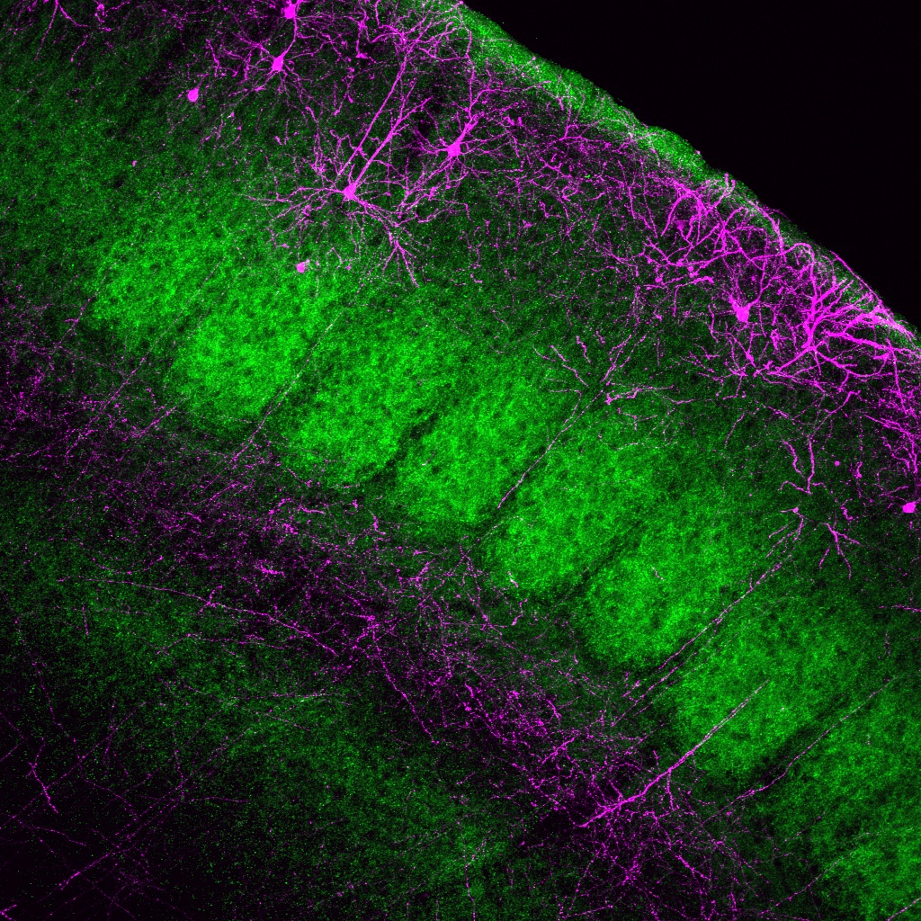
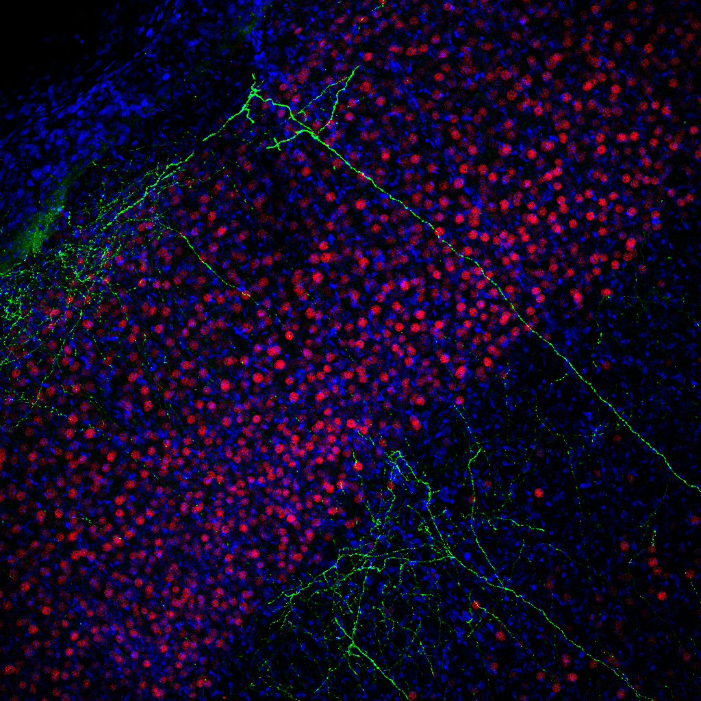
We have adapted monosynaptic pseudo-rabies tracing to quantitatively map the inputs received by specific pyramidal neurons in the mouse cortex using combination of sparse in utero cortical electroporation (IUCE) using a Cre- dependent expression FLEX-plasmid expressing TVA receptor, N2c Glycoprotein, and histone- tagged-GFP (hGFP). TVA expression enables infection by EnvA pseudotyped G-deleted N2c rabies virus (Rabv), while the N2c glycoprotein is required for trans- synaptic and retrograde spread of the virus. Hence, sparse Cre-induced expression of hGFP, TVA and N2c in a sparse population of layer 2/3 pyramidal neurons primes these ‘starter’ neurons for infection and trans- synaptic labeling by the pseudotype. Ewoud Schmidt in our lab also developed a computational pipeline to map the 3D position of all traced neurons from multiple mouse brains in a common reference Atlas (Allen Brain Atlas).
This work is featured in this paper by Ewoud Schmidt.

2-Photon Calcium Imaging in awake behaving mice
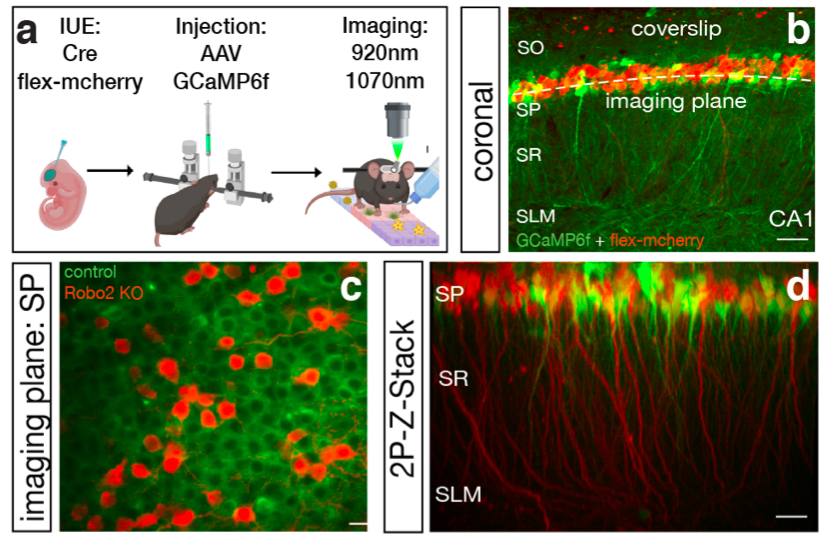
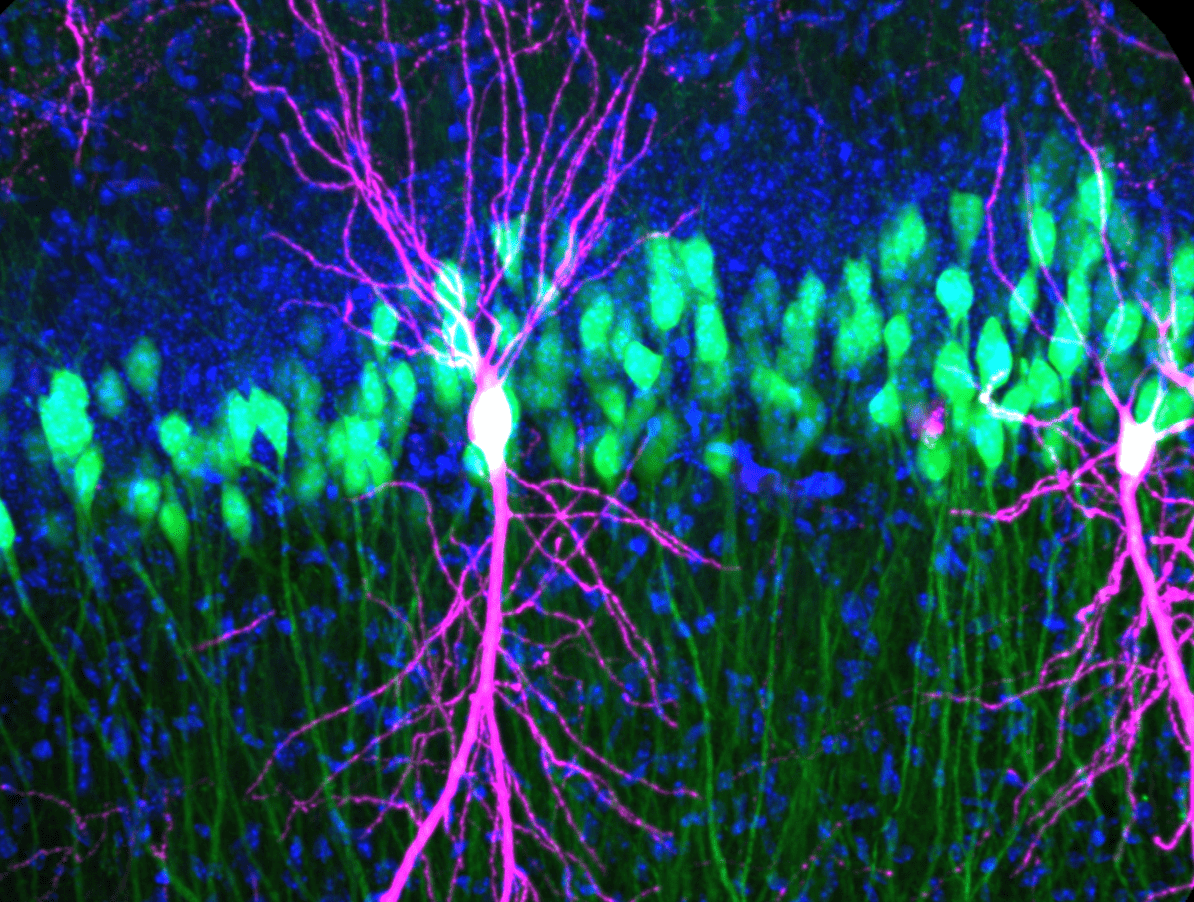
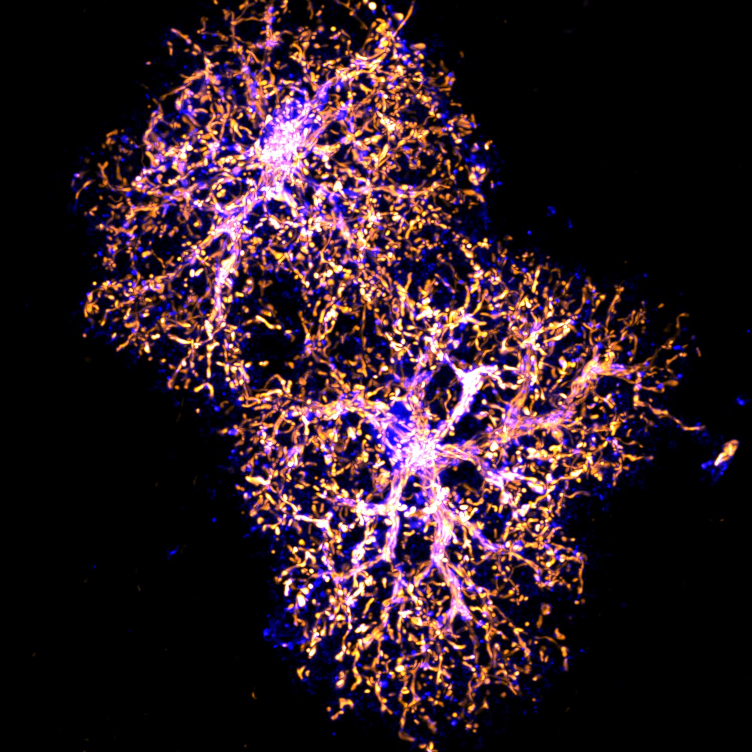
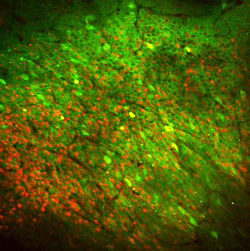
We implemented in vivo 2-photon (2P) Ca2+ imaging in awake behaving mice to probe either in cortical or hippocampal circuit function. For example, in close collaboration with our colleague in Attila Losonczy's lab , Heike Blockus has used this approach to probe changes in the response properties of control or Robo2-deficient in hippocampal CA1 pyramidal neurons in the same mice. She found that reduction by 40% of inputs from CA3 in Robo2-deficient CA1 PNs significantly alters the fraction of spatially tuned CA1 PNs and alters spatial coding. Ewoud Schmidt in our lab also used in vivo 2-photon Ca2+ imaging to demonstrate that humanization of SRGAP2C expression in layer 2/3 of the barrel cortex increases the reliability and selectivity of sensory evoked responses.
This work is featured in the latest papers of Ewoud Schmidt and Heike Blockus.

Publications and News

Funders

Zuckerman Mind Brain Behaviour Institute

Jerome L. Greene Science Center

Columbia University

New York